Atomic Relaxation in HPGe Crystals
The modeling process in the GEIM engine is divided into two distinct physical domains: high-energy interaction profiles with deep inner shells of an isolated germanium atom, and subsequent structural energy redistribution within the semiconductor crystal lattice.
Atomic Subshell Energy Levels
When individual germanium atoms bind to form the diamond cubic lattice of a High-Purity Germanium (HPGe) detector, the interatomic spacing stabilizes around 0.245 nm. At this scale, the inner core electronic shells (1s, 2s, 2p, 3s, 3p, 3d) remain highly localized and are effectively shielded from crystal field perturbations. However, the outermost valence orbitals (4s and 4p) undergo complete collectivization, morphing into delocalized energy bands.
Below is the engineered layout of germanium energy levels inside the crystal matrix, rendered utilizing a modified logarithmic scaling mechanism to display both deep-core bound states (11.1 keV) and subtle edge-band gaps on a single continuous chart.
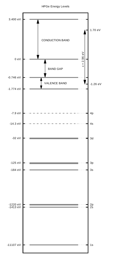 Figure 1: Band structure and atomic shell energy profiles within a germanium crystal. The 4s and 4p subshells are dashed to reflect their dissolution into the valence band.
The cumulative excess kinetic energy deposited by incoming primary particles, subsequent Auger electrons, and secondary cascades is balanced in the semiconductor via two competing relaxation channels: 1. Ionization Signal: Electron-hole pair generation via impact ionization, forcing valence-band electrons across the band gap (Δ E = 0.746 eV) into the conduction band. The average macroscopic energy required to create a stable charge-carrier pair is \(\epsilon \simeq 2.96\) eV. 2. Thermal Losses: Excitation of mechanical lattice vibrations (acoustic and optical phonon emission).
Transition Probability Matrices
When a primary core vacancy is induced via photoabsorption or electron impact, a rapid cascade of atomic relaxation is triggered. The vacant state is filled by an electron dropping down from a less tightly bound upper subshell.
To model this stochastic pathway via Monte Carlo algorithms, raw cross-sections from the EADL (Evaluated Atomic Data Library) are restructured into a two-step random walk formulation.
K-Shell ($\(1s_{1/2}\)$) Cascade Example
Below are the mapped transition profiles for a primary vacancy initialized at the deepest atomic core bound state.
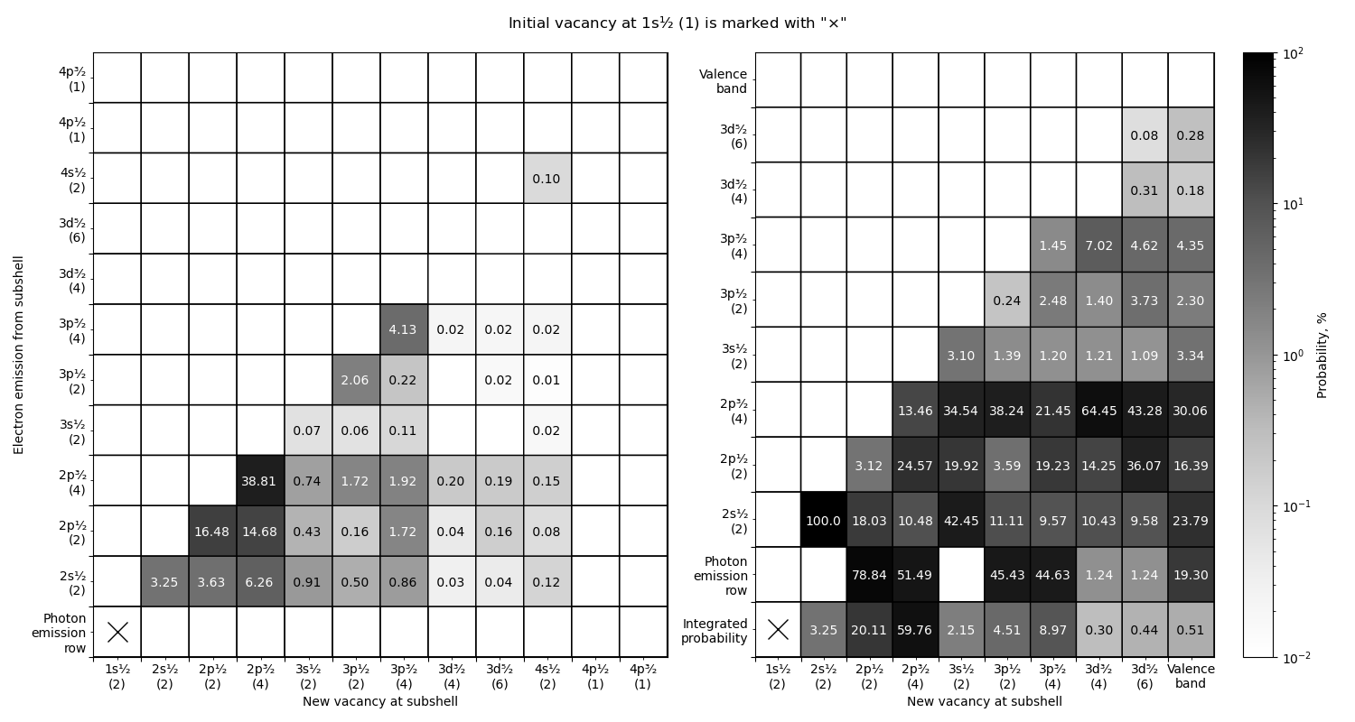
Figure 2: Statistical probability layout for a primary vacancy at the K-shell (marked with a black cross "×").
Structural Breakdown of the Maps:
-
Left Panel (Original EADL Records):
- Displays the global baseline of a single isolated step of vacancy filling.
- Photon Emission Row: Displays characteristic X-ray fluorescence pathways. For example, a \(2p_{3/2} \to 1s\) drop (\(K_{\alpha1}\) line) occurs with a probability of 38.81%, destroying the core vacancy and leaving a secondary vacancy at the \(2p_{3/2}\) level.
- Matrix Field: Non-radiative transitions (Auger and Coster-Kronig paths). The drop energy is collisionally transferred to another bound electron, ejecting it into the continuum. The horizontal coordinate denotes the first newly created vacancy, while the vertical axis tracks the shell from which the Auger electron was expelled (second vacancy). The global integral of the matrix equals 100%.
-
Right Panel (Restructured Monte Carlo Matrix):
- Optimized for lightning-fast discrete random sampling. All boundary valence configurations (4s, 4p) are mathematically collapsed into a unified Valence band node.
- Integrated Probability Row: The first phase of the random walk selection. Represents the absolute sum probability that the current vacancy will draw an electron from this specific column, regardless of the physical mode. The row sum matches 100%.
- Vertical Columns: The second phase of the random walk. Given a chosen column from step one, the vertical split stochastically determines the specific emission branch. The
Photon emission rowelement dictates the probability of a pure radiative photon release, while upper indices dictate the precise shell responsible for an Auger electron emission. Every isolated vertical column integrates strictly to 100%.